תיאור
Description
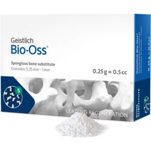
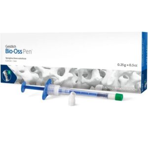
Geistlich Bio-Oss® Collagen consists of 90% Geistlich Bio-Oss® granules and 10% porcine collagen. The Geistlich Bio-Oss® particles provide Geistlich Bio-Oss® Collagen with all the advantages of the scientifically proven No 1. biomaterial in regenerative dentistry1.
The additional 10% of porcine collagen make it formable and easy to handle. Its regenerative potential clearly distinguishes Geistlich Bio-Oss® Collagen from mere collagen plugs.
Usage
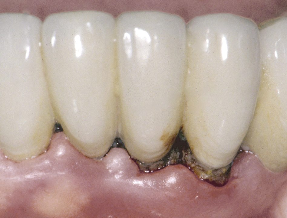
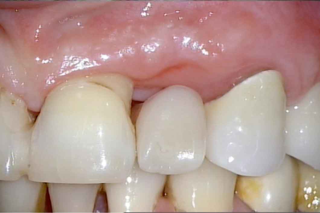
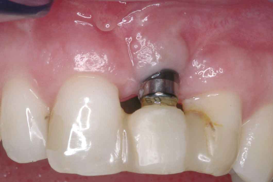
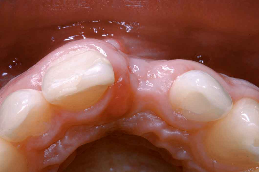
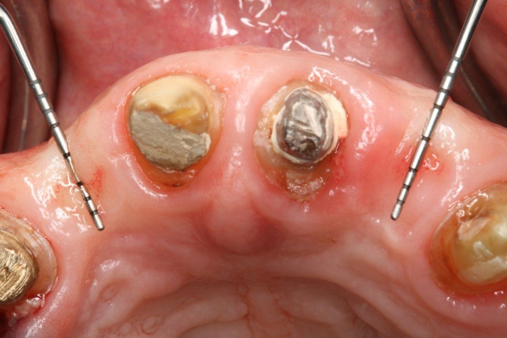
Geistlich Bio-Oss® Collagen is used in a variety of indications, including ridge preservation, minor bone augmentation and periodontal regeneration. The collagen is absorbed after a few weeks and does not replace the barrier function of a membrane.
Geistlich Bio-Oss® Collagen is “the Master’s Choice” because:
- The added collagen in Geistlich Bio-Oss® Collagen improves handling and tailoring to the morphology of the defect site2,3.
- Geistlich Bio-Oss® particles serve as a scaffold for new bone and ensure predictable bone regeneration4,5.
- Geistlich Bio-Oss® Collagen augmented tissue remains volume-stable in the long term due to the low resorption rate of the material6,7.
- Geistlich Bio-Oss® Collagen significantly improves clinical attachment and pocket depth in periodontal surgery8.
- Geistlich Bio-Oss® Collagen has the capacity to enable regeneration of the periodontal attachment apparatus in intrabony defects9.
- Handling characteristics are enhanced through the addition of 10% collagen. Geistlich Bio-Oss® Collagen is easily modelled and adheres well to the defect site.
References:
1. iData Research Inc., US Dental Bone Graft Substitutes and other Biomaterials Market, 2015 iData Research Inc., European Dental Bone Graft Substitutes and other Biomaterials Market, 2015.
2. Trevisiol L, et al.: J Craniofac Surg 2012, 23(5): 1343-48.
3. Rohner D, et al.: Int J Oral Maxillofac Surg 2013; 42(5): 585-91.
4. Cardaropoli D, et al.: Int J Periodontics Restorative Dent 2012, 32(4): 421-30.
5. Jung RE, et al.: J Clin Periodontol 2013, Jan; 40(1): 90-98.
6. Araujo MG, et al.: Clin Oral Implants Res 2010; 21(1): 55-64.
7. Mordenfeld AT, et al.: Clin Implant Dent Relat Res 2012, Oct 15 (Epub ahead of print).
8. Sculean A, et al.: J Clin Periodontol 2005: 32: 720-24.
9. Nevins ML, et al.: Int J Periodontics Restorative Dent 2003; 23: 9-17.